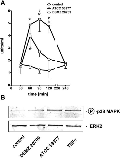
FIG. 5.
HUVEC were stimulated at an MOI of 10 for each strain. ELISA for phospho-p38 MAPK (for details, see Materials and Methods) revealed a time-dependent phosphorylation of p38 MAPK in ATCC 53977- and DSMZ 20709-treated endothelial cells (A). Strain ATCC 53977 induced a prolonged phosphorylation of p38 MAPK, while the signal in DSMZ 20709-stimulated cells decreased again after 60 min. Western blot analysis confirmed p38 MAPK phosphorylation 60 min postinfection in stimulated HUVEC (MOI of 10) (B). A 12.5% polyacrylamide gel was used; ERK2 served to confirm equal loading with proteins. TNF-α (10 ng/ml; 60 min) was used as a positive control. Data presented in panel A are the means ± SEM of three separate experiments. *, P < 0.05 compared to control; #, P < 0.05 comparing DSMZ 20709 versus ATCC 53977. In panel B one representative blot of three is shown.

