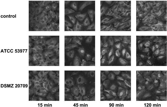
FIG. 7.
NF-κB activation in P. gingivalis-stimulated HUVEC was analyzed by using immunofluorescence. HUVEC grown on Thermanox slides were stimulated with both strains for the times indicated. Cells were fixed and permeabilized as indicated in Materials and Methods, incubated with a polyclonal rabbit anti-human NF-κB p65 antibody, followed by the addition of an Alexa Fluor 488-conjugated goat anti-rabbit Ig antibody. The top row shows mock-treated control cells. Note a time-dependent increasing fluorescence intensity in the nuclei of P. gingivalis-stimulated HUVEC, demonstrating the nuclear translocation of NF-κB starting after 45 (ATCC 53977) to 90 min (DSMZ 20709) postinfection. Representative pictures (of three independent experiments; magnification, ×640) are shown.

