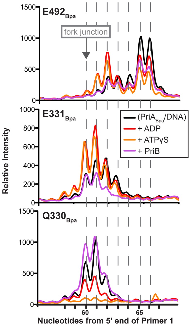
Figure 6.

Nucleotide and PriB alter PriABpa–DNA fork crosslinks. Densitometry profiles of nucleotide crosslink sites on the template lagging strand, from primer extension gels as in Figure 5B, for PriA variants with Bpa incorporated at E491 (top), E330 (middle) and Q329 (bottom), when crosslinks are formed in the presence of no additional factors, ADP, ATPγS or PriB. Data are quantified from a representative gel of greater than three replicates.
