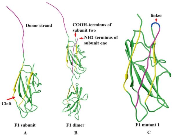
Fig. 1.
Reorientation of the NH2-terminal β-strand of F1 protein to generate monomeric F1. (a) Structure of F1 subunit, purple strand indicates donor β-strand, yellow strands indicate the strands that form groove. (b) F1 dimer showing how F1 monomers oligomerize to generate a linear fiber. (c) Schematic of F1 mutant 1 in which the NH2-terminal β-strand is reoriented

