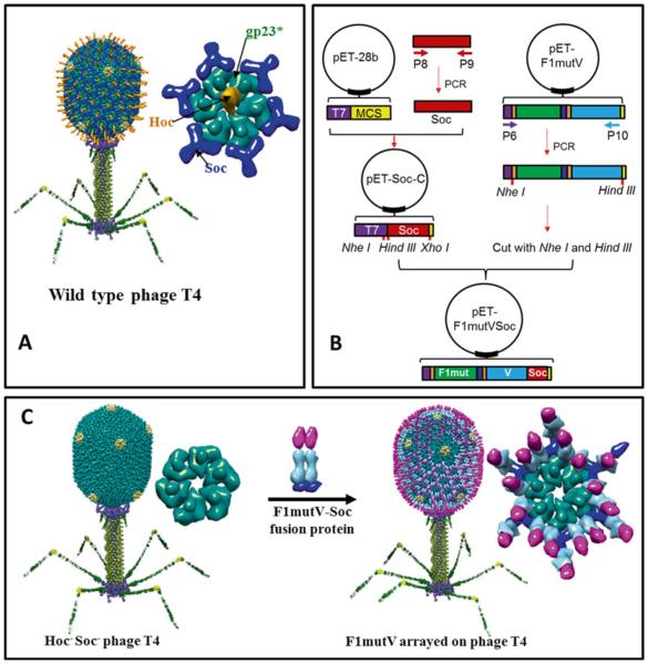
Fig. 3.
Preparation of T4 nanoparticle-arrayed plague vaccine. (a) Structural model of bacteriophage T4. The enlarged capsomer on right shows the major capsid protein gp23* (green; “*” represents the cleaved mature form) (930 copies), Soc (blue; 870 copies), and Hoc (orange; 155 copies). Yellow subunits at the fivefold vertices correspond to gp24*. The portal vertex (not visible in the picture) connects the head to the tail. (b) Cloning strategy to generate pET-F1mutVSoc. Red indicates Soc. The other components are shown as in Fig. 2. P6–P10 indicate primers 6–10. (c) Display of F1mutV-Soc fusion protein on the Hoc−Soc− phage particle. Shown on the right are models of the enlarged capsomers before and after F1mutV-Soc display

