FIG. 6.
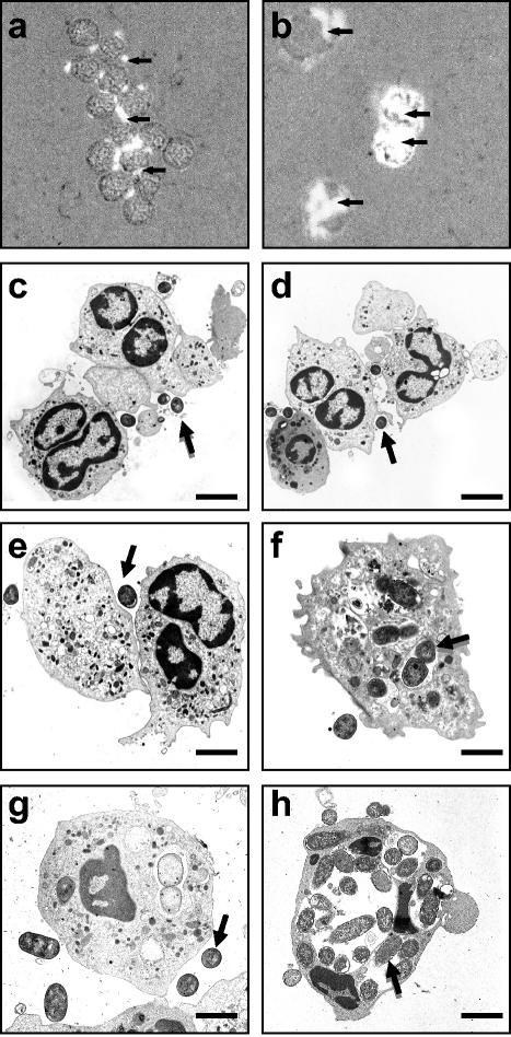
(a and b) Light microscopic analysis of PMNs infected with GFP-IH11128 (a) and GFP-DH5α (b) E. coli strains. The bacteria were left in contact with the PMNs for 1 h. Magnification, ×400. The arrows indicate GFP bacteria. (c to h) Transmission electron micrographs of nontransmigrated (c to f) and transmigrated (g and h) PMNs infected for 1 h with E. coli strains IH11128 (c and g), pSSS1-DH5α (d), C1845 (e), and DH5α (f and h). The arrows indicate bacteria. Bars: 10 μm (c and d) and 2 μm (e to h). The results are representative of three experiments performed on different PMN preparations.

