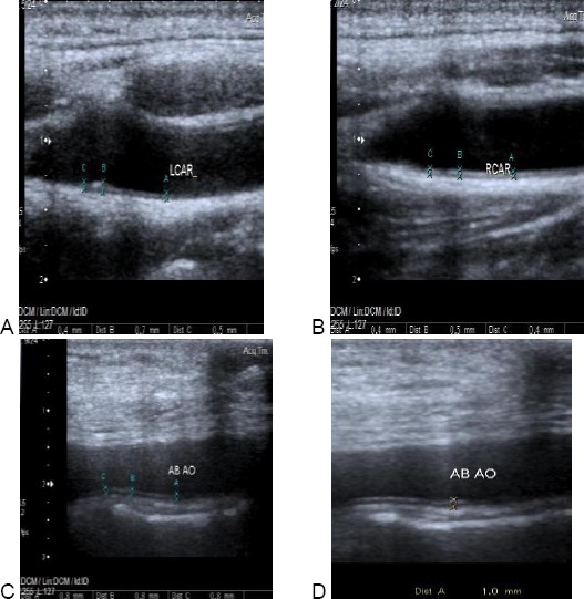
Figure 3.

Carotid & aortic intimal medial thickness in one of the patients. A: showing normal Rt. cIMT of one of the patients. cIMT = 0.4-0.5 mm. B: showing increased Lt. cIMT of the same patients. cIMT = 0.4-0.7 mm. C &D: showing increased aIMT of the same patient. aIMT = 0.8-1 mm
