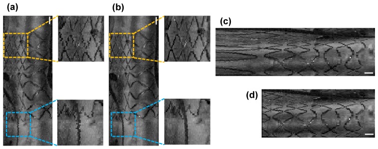
Fig. 5.
(a) An en face projection of a stented coronary segment before NURD correction in the ECG-triggered high speed OCT imaging (500 rps). Stent structure (yellow dashed box) and a guidewire (blue dashed box) are shown as if they are in zigzag shapes. (b) An en face projection of the same stented coronary segment after NURD correction. Stent structure (yellow dashed box) and a guidewire (blue dashed box) are smoothly connected. (c) An en face projection before non-uniform pullback distortion correction. (d) An en face projection after non-uniform pullback distortion correction. Scale bars, 1 mm.

