Fig. 5.
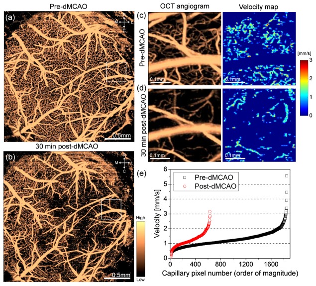
OCT capillary velocimetry in ischemic stroke model of mouse in vivo. OCT angiograms of the mouse cortex (surface to cortical layers II/III) through cranial window before (a) and 30min after (b) distal middle cerebral artery occlusion (dMCAO). Rarefaction of perfused capillaries is apparent after occlusion compared to pre-dMCAO. (c,d) OCT angiograms (boxes in (a) and (b)) (left) and corresponding capillary velocity maps (cortical layers II/III) (right) before (c) and after (d) occlusion, respectively. The velocity map post-dMCAO (d) shows reduction in velocity in the local perfused capillaries compared to pre-dMCAO (c). (e) Velocities in the capillary velocity maps before and after occlusion, plotted against the magnitude. For both pre/post-dMCAO, the RBC velocities are not constant but variable, clustered at around 1.0mm/s, but smearing at higher velocities up to 5.5mm/s before occlusion. R: rostral, C: caudal, M: medial, L: lateral.

