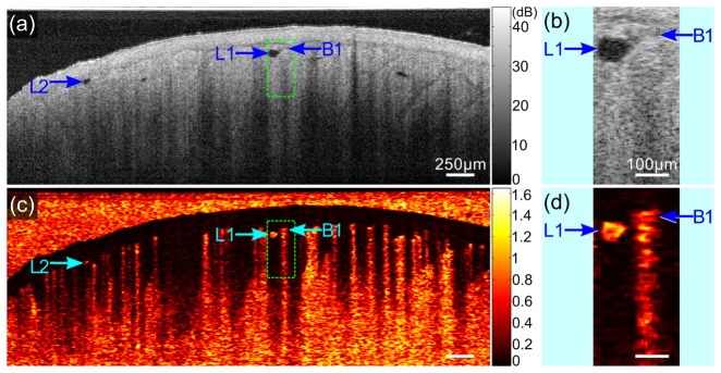
Fig. 2.
Characteristic of lymphatic vessels in speckle decorrelation. (a) and (c) Cross-sectional image of the raw OCT signal and the resulting speckle decorrelation of the scar tissue in Fig. 1. (b) and (d) Magnified images of the regions in the dashed rectangles in (a) and (c). L1 and L2: lymphatic vessels; B1: blood vessel. Scale bars in (a) and (c): 250 µm; (b) and (d): 100 µm.

