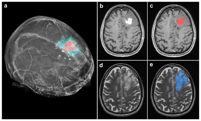
Fig. 5.
(a) 3-Dimensional volume rendering from the preoperative MRI of a patient with a grade 4 glioblastoma. The tumor regions visible on T1- and T2-weighted MRI scans are indicated in red and blue respectively. White ‘plus’ symbols indicate measurement locations of invasive cancer detected with RS. (b) A T1-weighted MR axial image with the glioblastoma visible. (c) The same image as in b, with the segmentation of the tumor region visible in red. (d) A T2-weighted MR axial image with the glioblastoma visible, corresponding to the same cross section as in b and c. (e) The same image as in d, with the segmentation of the tumor region visible in blue.

