
Figure 4.
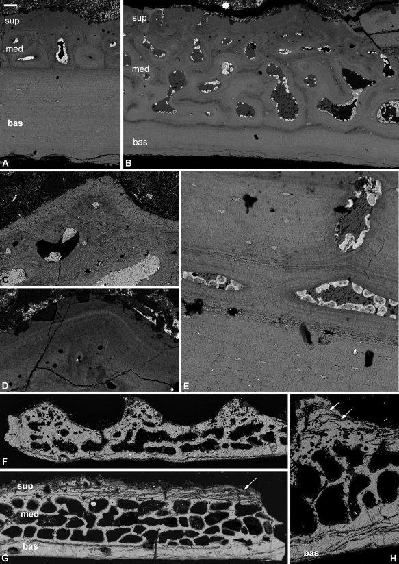
Histology of the phyllolepid arthrodire Phyllolepis (A–E) and the petalichthyid Lunaspis (F–H). Phyllolepis anterior lateral plate NHMUK PV P.50928: three‐layered structure with large basal layer (A), and large medial layer with cancellar spaces centripetally infilled (B), detail of the superficial layer with capping enameloid layer (C) and buried asymptotically tapering enameloid layer (D), section through a scarf joint basal layer with Sharpey's fibers and cell lacunae (E). Lunaspis ventrolateral plate ANSP 21405: three‐layered structure in peripheral, thinner part of the plate (F), three‐layered structure in thicker part of the plate with larger medial layer and parallel cracks in the superficial layer probably reflecting lamellae (G), detail of a tubercle showing cracks reflecting lamellae (H). bas, basal lamellar layer; en, enameloid; med, medial layer; sup, superficial layer. Scale bar equals 50 μm in (A), 67 μm in (B), 26 μm in (C), 30 μm in (D), 20 μm in (E), 157 μm in (F), 150 μm in (G), and 75 μm in (H).