
Figure 1.
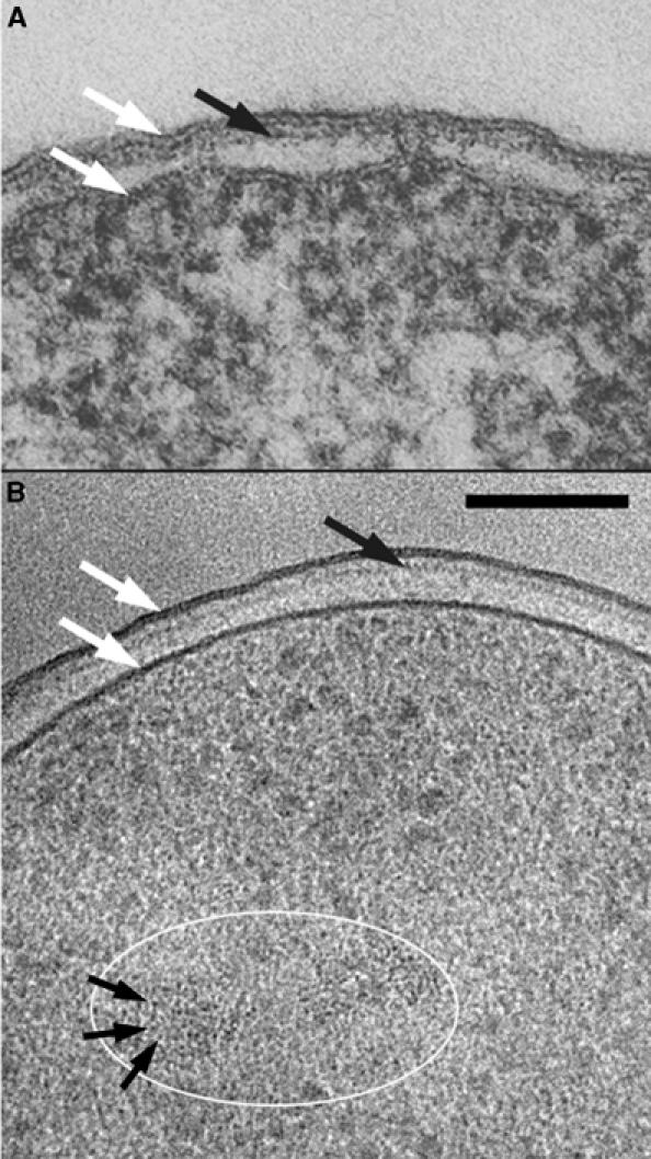
Gram-negative bacteria. (A) Escherichia coli K12 embedded in conventional resin (from Beveridge, 1999, Figure 1, with permission). (B) Pseudomonas aeruginosa PAO1 prepared by CEMOVIS (Matias et al, 2003). The cells in exponential growth were put in 20% (wt/vol) dextran (42 kDa) and vitrified in a Leica EMPACT (Vienna, Austria) high-pressure freezer. The sections were cut at −160°C in a Leica UCS/FCS cryomicrotome with a 45° Diatome diamond cryo-knife (Biel, Switzerland). Observation was made at −180°C in a Philips CM12 EM (Eindhoven, The Netherlands) operating at 80 kV and equipped with a Gatan 626 cryo-specimen holder (Warrendale, PA) and a Gatan 600CW Multiscan CCD camera. The total electron dose did not exceed 800 e/nm2. Technical details are described in previous publications (Dubochet et al, 1988; Al-Amoudi et al, 2002) or are in preparation. White arrows: cell membrane and outer membrane; black arrows: peptidoglycan layer; the white ellipse marks a region of the nucleoid; small black arrows: 2 nm high-contrast dots identified as portions of DNA filaments seen along their axis. Scale bar: 100 nm.