
Figure 2.
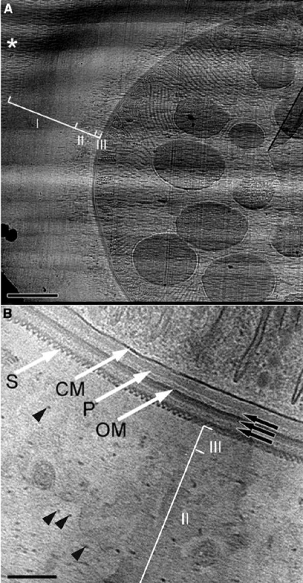
Cyanobacterium L. majuscula prepared by CEMOVIS. (A) Low-magnification view. I, II and III are different zones of the extracellular matrix; white asterisk: bulk medium with crevasses. Scale bar: 1 μm. (B) High-magnification view of the envelope. CM: cell membrane; P: peptidoglycan; OM: outer membrane; S: putative S-layer; black arrows: additional layers; II and III: zones of the extracellular matrix; black arrowheads: filaments in zone II. Scale bar: 100 nm.