
Figure 3.
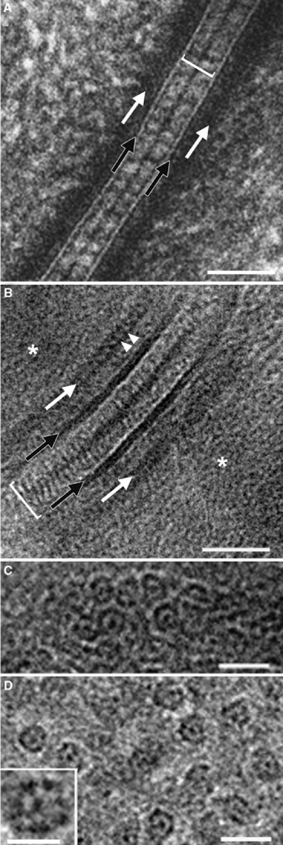
Epidermal desmosome and IFs. (A) Desmosome from neonatal mouse epidermis prepared by freeze substitution (from He et al, 2003, from Figure 1B, with permission). (B) Human desmosome at midportion of the viable part of forearm epidermis prepared by CEMOVIS. A ca. 200 μm thick layer was sliced with a razor blade and immediately cooled in a Baltec HPM 010 (Balzers, Liechtenstein) high-pressure freezing apparatus. The rest of the preparation was as for Figure 2B. See text for more explanations. (C) Same region as in (B) but with IFs oriented along the viewing direction. (D) As in (C) but in stratum corneum. The fine structure of the IFs is best resolved in the thinnest sections (inset). (C, D) Modified from Norlén and Al-Amoudi (2004), with permission. Section thickness: 70 nm (B, C); less than 50 nm (D). Scale bar: 50 nm (A, B); 20 nm (C, D); 10 nm (inset, D).