
Figure 4.
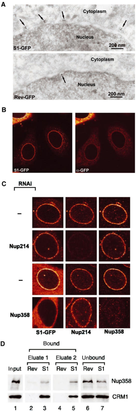
S1 NES localises at Nup358. (A) Immunoelectron microscopy. Cryosections of MCF-7 cells transfected with S1-GFP- or RevNES-GFP-containing reporter constructs (see Figure 2) were labelled with anti-GFP antibodies followed by protein A gold. In cells expressing S1-GFP, protein gold (arrows) decorates the outer aspect of the nuclear envelope at NPCs. (B) Immunofluorescence. Cells as in (A) were permeabilised with low concentrations of digitonin such that the nuclear membrane remained intact and labelled with anti-GFP antibodies. Anti-GFP antibodies stain the NE and colocalise largely with the signal from GFP. (C) Knockdown of Nup358 by RNAi removes S1-GFP from the NE. HeLa cells were cotransfected with a plasmid expressing shRNAs targeting Nup358 or Nup214 and an S1-GFP-containing reporter plasmid. Cells were analysed 72 h post-transfection for Nup358 and Nup214 levels and S1-GFP by indirect immunofluorescence and direct GFP fluorescence, respectively. A strong knockdown of Nup358, but not of Nup214, reduces S1-GFP from the NPC. (D) S1 NES physically interacts with Nup358. Proteins from Xenopus interphase egg extracts were affinity selected on immobilised biotinylated Rev or S1 NES peptides. Starting material (lane 1) and bound (lanes 2–5) and unbound (lanes 6 and 7) fractions were analysed for the presence of Nup358 and CRM1 by Western blotting.