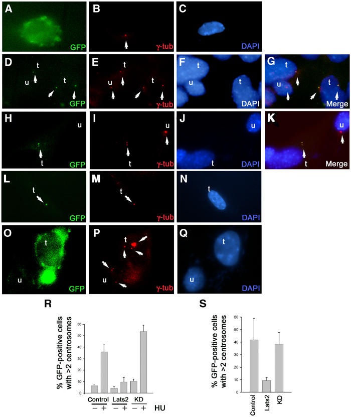
Figure 7.

Lats2 is a centrosomal protein that suppresses centrosome overduplication. (A–K) Subcellular localization of GFP-Lats2 at centrosomes by indirect immunofluorescence. Wild-type MEFs transfected with either GFP (A–C), GFP-Lats2 (D–K) or GFP-Lats2KD (L–N) expression constructs were analysed 24 h later for GFP fluorescence (A, D, H, L) and γ-tubulin (red) immunofluorescence (B, E, I, M), and DNA was stained with DAPI (C, F, J, N). Merged images G and K confirming colocalization of GFP-Lats2 and γ-tubulin are derived from (D–F) and (H–J), respectively. Transfected cells (GFP-positive) and untransfected cells in (D–N) are denoted as t and u respectively; arrows indicate centrosomes (B, E, I, M, P), GFP-Lats2 fluorescence (D, H), GFP-Lats2KD fluorescence (L) and merged signals (G, K). (O–Q) Centrosome overduplication in the presence of HU is suppressed by GFP-Lats2. Representative GFP vector-transfected cell (t) and untransfected (u) cell (O) demonstrating supernumerary centrosomes (arrows) as visualized by γ-tubulin immunostaining (P). DNA is visualized by DAPI stain (Q). (R) Graph depicting the percentage of GFP-positive cells exhibiting >2 centrosomes from wild-type MEFs transfected with GFP vector, GFP-Lats2 (Lats2) or GFP-LatsKD (KD) in the absence or presence of 2 mM HU. (S) Centrosome overduplication in Lats2-deficient MEFs is suppressed by GFP-Lats2. Graph depicting the percentage of GFP-positive cells exhibiting >2 centrosomes from Lats2−/− MEFs transfected with GFP (control), GFP-Lats2 (Lats2) or GFP-Lats2KD (KD).
