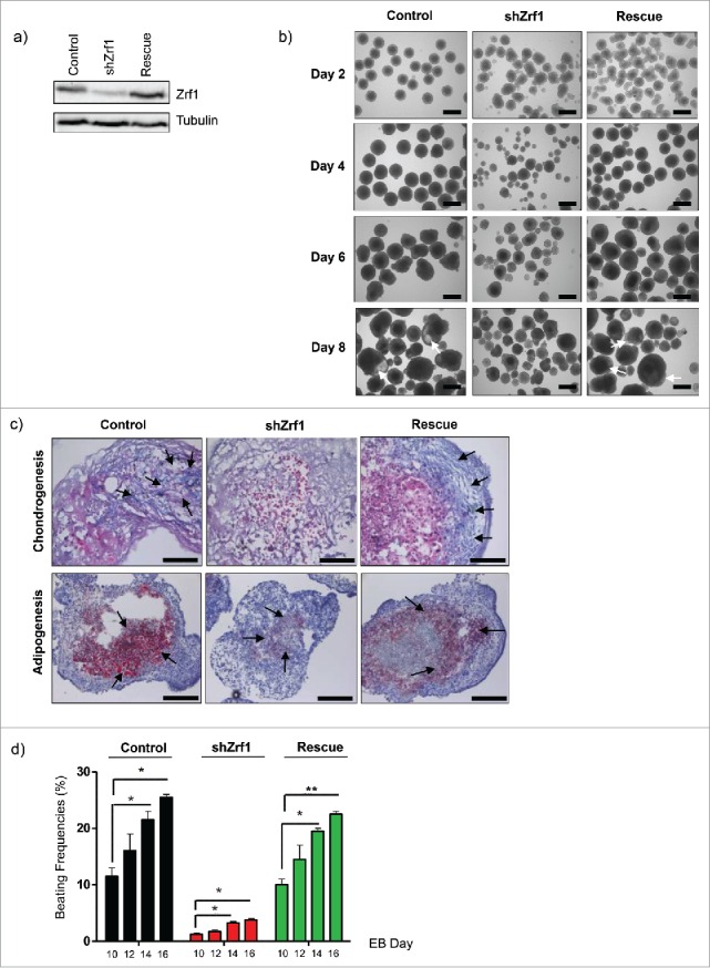
Figure 2.

Restoration of Zrf1 expression in Zrf1 knockdown ES cells rescues the mesoderm phenotype. (A) Western blot analyzing Zrf1 levels in control, shZrf1 and rescue cells. Alpha tubulin was used as a loading control. (B) Brightfield images of control, shZrf1 and rescue cells derived EBs were taken at 4x magnification at days 0, 4, 6 and 8. White arrows indicate the cystic cavities in control and rescue cell derived EBs. Scale bars, 500 µm. (C) Representative images of alcian blue (upper panel) and oil-red-O (lower panel) stainings of control, Zrf1 knockdown and rescue EBs were taken at either 40x or 20x magnification after 16 days of differentiation. Black arrows indicate mesoderm derived tissues in alcian blue staining. Cartilage: blue, purple. Bone marrow: dark blue. Black arrows indicate mesoderm derived lipid droplets in oil-red-O staining. Scale bars, upper panel 50 µm, lower panel 100 µm. (D) The numbers of spontaneously beating EBs were counted under an inverted-light microscope on Day 10, 12, 14 and 16 of the culture. Data represent the average of 3 experiments, +/− SEM *p < 0.5, **p < 0.01, as calculated by 2-tailed unpaired t test.
