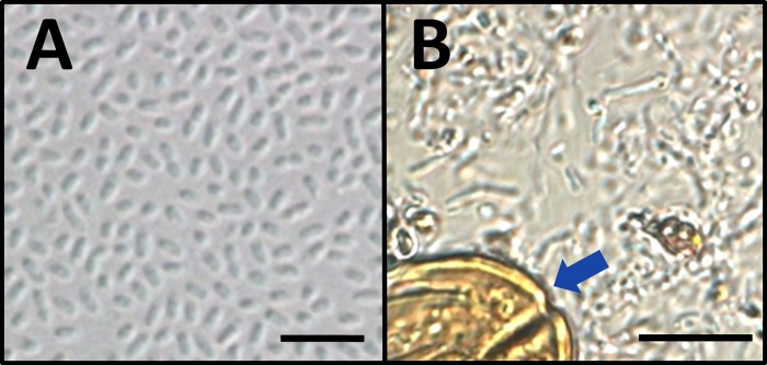
Fig 2. Photomicrographs representing direct wet mount images of hemolymph from SIW.
Panel A shows a hemolymph sample obtained from one SIW, including a high density of bacteria with uniform morphology (Ss1), and an absence of pollen grains. Panel B shows a wet mount of hemolymph that was obtained from a different SIW, revealing mixed bacterial morphologies and occasional pollen grains (blue arrow). Cultures prepared from SIW collectively grew >1.0e9 cfu/ml Ss1. The scale bar represents 5 μm in Panel A and10 μm in Panel B.

