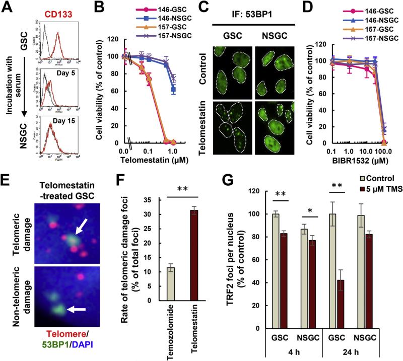
Fig. 1.
Telomestatin induces telomere dysfunction and inhibits GSC growth. (A) Measurement of CD133 levels in GBM146 cells by flow cytometry. Black histogram indicates normal immunoglobulin as negative control. (B) Effect of telomestatin on growth of GSCs and NSGCs. GBM146 and GBM157 cells were treated with telomestatin for 144 h. Error bar, standard deviation. (C) DNA damage foci induced by telomestatin in GSCs. GBM146 cells were treated with 5 μM telomestatin in serum-free medium for 24 h and subjected to immunofluorescence staining with anti-53BP1 antibody. (D) Effect of telomerase inhibitor BIBR1532 on growth of GSCs and NSGCs. GBM146 and GBM157 cells were treated with indicated concentrations of BIBR1532 for 144 h (E) iFISH assay. GSCs were treated with 1 μM telomestatin in serum-free medium for 96 h. Representative images of telomeric and non-telomeric DNA damage foci are shown (arrows in upper and lower panels, respectively). Red, telomere; green, 53BP1; blue, DAPI staining for nuclear DNA. (F) The rate of telomeric 53BP1 damage foci in telomestatin- or temozolomide-treated GSCs. GSCs were treated with 1 μM telomestatin or 10 μM temozolomide in serum-free medium for 96 h (these drug concentrations inhibited the cell growth to equivalent extents: data not shown). The rate of telomeric 53BP1 foci among all 53BP1 foci was calculated. (G) TRF2 immunofluorescence staining. GBM146 cells were treated with 5 μM telomestatin in serum-free medium for 4 or 24 h. The numbers of TRF2 foci per nucleus were counted and normalized against those in GSC/control cells. Statistical evaluations were performed using the Welch t-test. *, P < 0.05; **, P < 0.01. (For interpretation of the references to colour in this figure legend, the reader is referred to the web version of this article.)

