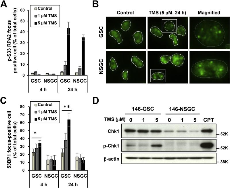
Fig. 4.
GSCs are sensitive to telomestatin-induced replication stress. (A) Immunofluorescence staining of telomestatin-treated GSCs and NSGCs with anti-p-RPA2-Ser33 antibody. GBM146 cells were treated with 1 or 5 μM telomestatin in serum-free medium for 4 or 24 h. Cells were classified as focus-positive or -negative according to numbers of punctate nuclear foci of p-RPA2-Ser33 staining (n > 3). (B) Representative image of A. (C) Immunofluorescence of telomestatin-induced 53BP1 foci in GSCs and NSGCs. GBM146 cells were treated as in A and classified according to the numbers of punctate nuclear 53BP1 foci (n > 4). Statistical evaluations were performed using the Welch t-test. *, P < 0.05; **, P < 0.01. (D) Western blot analysis. GBM146 cells were treated with 1 or 5 μM telomestatin for 24 h. Cell lysates were prepared and subjected to western blot analysis. CPT, GSCs were treated with 2 μM camptothecin for 1 h as positive control of p-Chk1.

