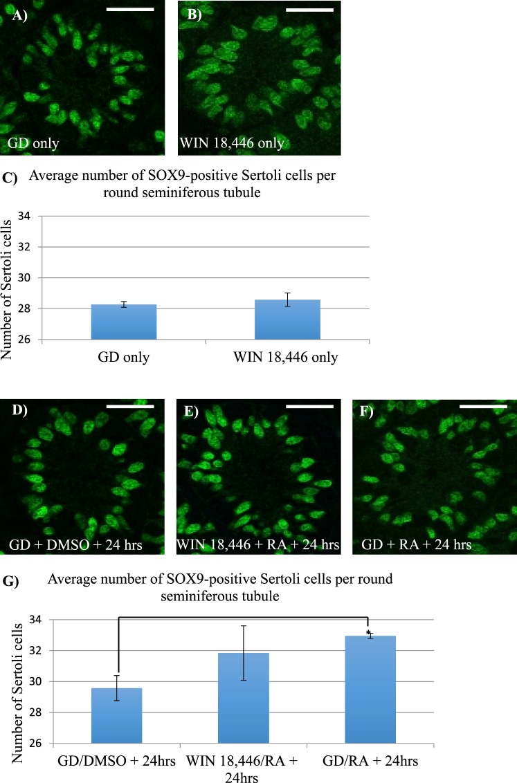
FIG. 9.
Excess RA exposure slightly increased the number of Sertoli cells in the postnatal testis. Immunofluorescent images depict representative cross sections of testes from mice treated with the WIN 18,446 vehicle control only (GD) (A), WIN 18,446-only (B), GD followed by RA vehicle (DMSO) (D), WIN 18,446 followed by an injection of RA (E), and GD followed by an injection of RA (F) stained for SOX9 expression. Bar graphs indicate the average number of SOX9-positive Sertoli cells per round tubule from testes of mice treated with either WIN 18,446-only compared to GD-only, displayed in C, or from mice treated with either WIN 18,446 followed by an injection of RA or GD followed by an injection of RA compared to GD followed by an injection of DMSO, displayed in G. * denotes a P-value = 0.01. Error bars represent SEM. White bars = 25 μm.

