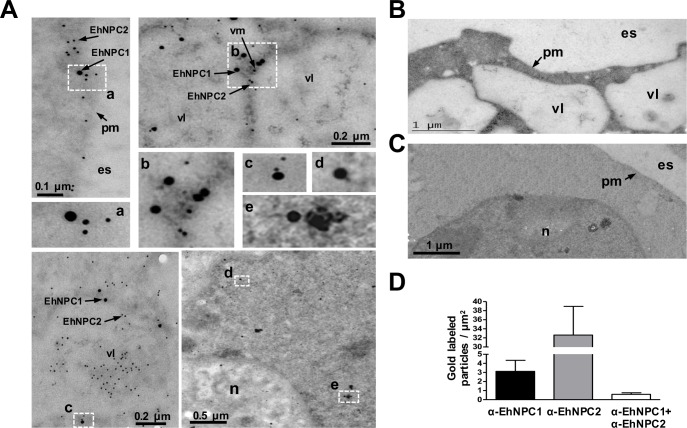
Fig 4. Localization of EhNPC1 and EhNPC2 in trophozoites analyzed by TEM.
(A) Thin sections of trophozoites were incubated with rabbit α-EhNPC1 and rat α-EhNPC2 antibodies, followed by incubation with gold labeled α-rabbit and α-rat secondary antibodies (20 and 10 nm gold particles, respectively). Squares indicate the magnified areas marked with the corresponding lower case letters. pm: plasma membrane, vl: vesicle lumen, vm: vesicle membrane, es: extracellular space, n: nucleus. (B, C) Controls using only secondary antibodies. (D) Graph showing number of EhNPC1 and EhNPC2 molecules recognized by the respective gold-labeled antibodies and their co-localization.

