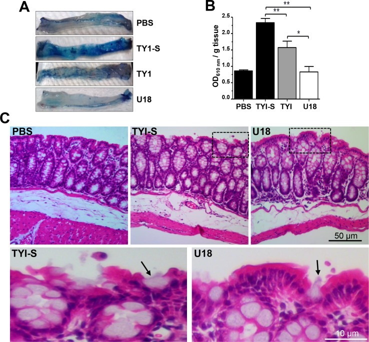
Fig 11. In vivo virulence of trophozoites cultured in TYI-S, TYI and TYI plus U18.
Capability of trophozoites to impair the mouse intestinal barrier. (A) Distal parts of the mouse colons after treatment with trophozoites or with PBS. Intestinal barrier impairment was measured as the ability of the Evans blue dye to permeate the mouse intestinal epithelium after contact with trophozoites. n = 5. (B) Data represent the mean ± standard error. PBS: mice undergoing surgery, but not inoculated with trophozoites. * p<0.05, ** p<0.01. (C) Hematoxylin-eosin staining of tissues. Squares were magnified in the corresponding lower panels. Arrows: trophozoites.

