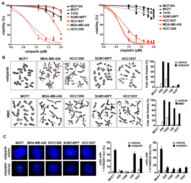
Figure 4. Characterization of BRCA1-mutated TNBC cell lines.
(A) Panel of indicated cell lines was treated with the indicated concentrations of veliparib or cisplatin and viability assessed after 7 days of treatment. (B) Cells were treated with vehicle, veliparib or mitomycin C and metaphase spreads were prepared; radials quantified in vehicle and drug-treated cells. (C) Cells were treated with vehicle, veliparib or cisplatin and analyzed by immunofluorescence for RAD51 and FANCD2 foci. Graphs show quantification of cells with > 5 foci in vehicle and drug-treated cells.

