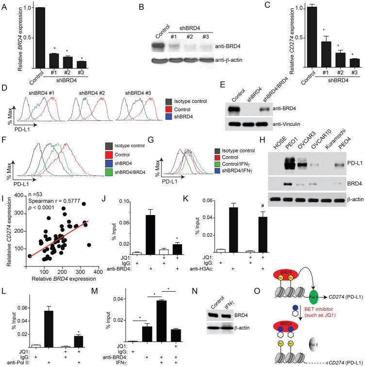
Figure 3. CD274 is a direct target gene of BRD4. See also Figure S3 and Table S2.
(A) PEO1 cells expressing shBRD4 or control were examined for BRD4 expression by qRT-PCR. * p<0.05.
(B) Same as (A), but examined for the expression of BRD4 protein by immunoblotting.
(C) Same as (A), but examined for CD274 mRNA expression by qRT-PCR. * p<0.05.
(D) Same as (A). PD-L1 expression was determined by FACS.
(E) PEO1 cells expressing shBRD4 (#3) that targets the 3′ UTR region of the human BRD4 gene with or without simultaneous expression of a wild-type BRD4 open reading frame. BRD4 and Vinculin expression was determined by immunoblot.
(F) Same as (E), but examined for PD-L1 expression by FACS.
(G) PEO1 cells expressing shBRD4 (#3) or control with or without IFNγ (20ng/L, 24h) stimulation were examined for PD-L1 expression by FACS.
(H) Expression of BRD4, PD-L1 and β-actin were examined in the indicated ovarian cancer cell lines or normal human ovarian surface epithelial cells by immunoblot.
(I) Correlation between BRD4 and CD274 expression was determined by Spearman statistical analysis in 53 cases of laser capture and microdissected high-grade serous ovarian cancer specimens.
(J-L) PEO1 cells were treated with or without 200 nM JQ1 for 24 hours. The cells were subjected to ChIP analysis using antibodies against: BRD4 (J), H3Ac (K), or Pol II (L). An isotype matched IgG was used as a negative control. The association with the CD274 gene promoter was quantified by qPCR. # p>0.05 and * p<0.05.
(M) Same as (J), but for PEO1 cells with or without IFNγ (20ng/mL, 24h) stimulation were treated with JQ1. * p<0.05.
(N) Same as (M), but examined for BRD4 and β-actin protein expression by immunoblot.
(O) A model for BRD4-mediated regulation of PD-L1 expression. Error bars represent S.E.M. of three independent experiments.

