
Figure 2.
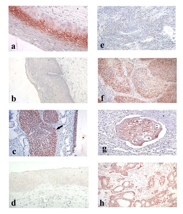
Cervical tissue samples after immunohistochemical staining with p16INK4a-specific antibodies. a. CIN I. Focal staining. b. CIN II. Negative staining. c. Cancer in situ (indicated with an arrow) and CIN III. Diffuse staining. d. CIN III. Negative staining. e. Squamous cell carcinoma. Negative staining. f. Squamous cell carcinoma. Diffuse staining. g. Squamous cell carcinoma embol. Diffuse staining. h. Adenocarcinoma. Diffuse staining.