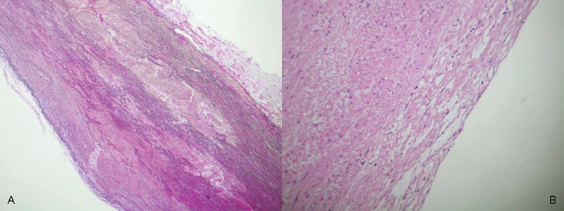
Fig. 3.

Histological examination of the patients' aortic wall (EvG staining, 100-fold magnification (A) and HE staining 200-fold magnification (B), respectively). pathological findings results in mucoid degeneration of the aorta (A) with lipidosis of the intimal layer (B).
