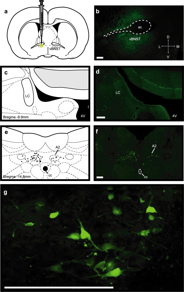
Figure 5.
Unilateral fluorogold tracing in the vBNST. (a,b) Schematic and representative infusion site of fluorogold into the vBNST. (c,d) Apparent lack of fluorogold-positive cells in the ipsilateral LC and atlas section corresponding to the photomicrograph. (e,f) Bilateral fluorogold-positive cells in the A2 and camera lucida drawing of labeled cells in the atlas section corresponding to the photomicrograph. (g) Higher magnification image of fluorogold-positive cells in the ipsilateral A2. Scale bars are 200 μm. 4 V, fourth ventricle; ac, anterior commissure; cc, central canal.

