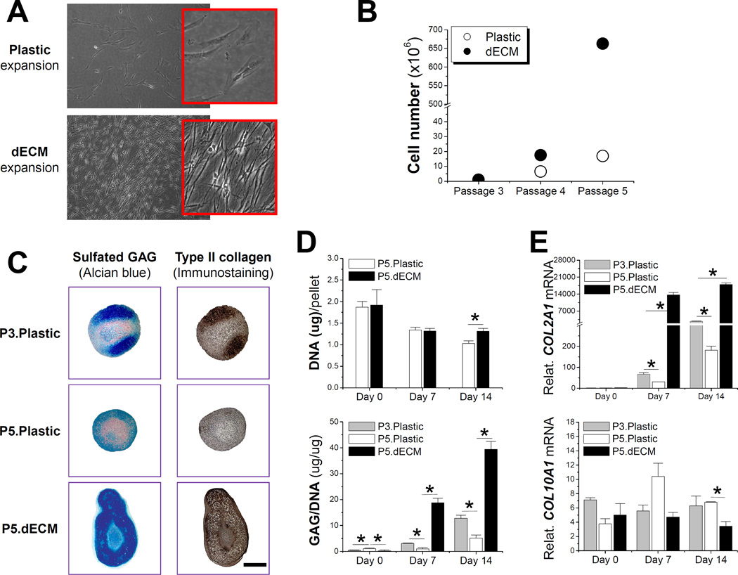
Fig. 1.
Effect of decellularized extracellular matrix (dECM) deposited by synovium-derived stem cells (SDSCs) on porcine SDSCs’ proliferation and chondrogenic differentiation. (A) Cell morphology five days after expansion on dECM and Plastic flasks; (B) Cell proliferation from passage 3 (P3) SDSCs grown on either dECM or Plastic flasks for two consecutive passages; (C) Alcian blue staining for sulfated GAG and immunostaining for type II collagen (scale bar: 800 mm) of 14-day chondrogenically induced SDSCs in a pellet culture system after two passages on dECM (P5.dECM) or Plastic flasks (P5.Plastic) with pre-expansion SDSCs (P3.Plastic) as a control; (D) Biochemical analyses were used to detect DNA content per pellet and ratio of GAG to DNA; (E) TaqMan real-time polymerase chain reaction (PCR) was used to quantitatively assess chondrogenic markers - COL2A1 (type II collagen) and COL10A1 (type X collagen). * indicates a statistical difference (p<0.05). Data are shown as average ± SD for n=6 in biochemical analyses and n=5 in real-time PCR. Reprint with permission from He, F.; Chen, X.; Pei, M. Tissue Eng. Part A 2009, 15, 3809. Copyright (2009) Mary Ann Liebert, Inc. Publications.

