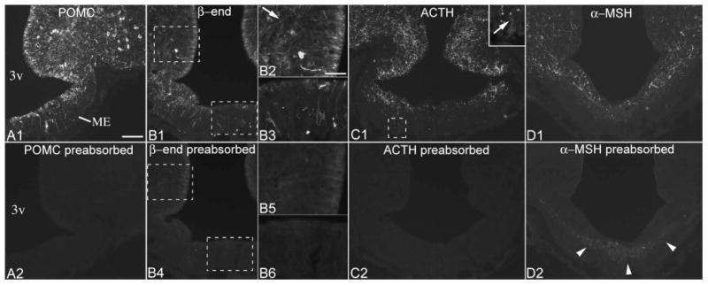
Figure 1.

Preabsorption controls for POMC, β-endorphin, ACTH and α-MSH antisera. (A1, A2) Adjacent sections from a brain with high POMC levels in tanycytes demonstrates that preincubation of the POMC antiserum with the immunizing peptide abolishes all POMC immunolabeling in tanycytes and neurons. (B1-B6) Preabsorption control for the β-endorphin antiserum in adjacent sections from a brain with intermediate POMC levels in tanycytes. Specific labeling is present in α2 tanycyte cell bodies and processes (arrow, B2), and in β2 and γ tanycytes (B3). In the section reacted with the preabsorbed antiserum (B4-6), some background labeling remains in the location of α2 tanycyte cell bodies, but this signal is indistinct. (C1, C2) Preabsorption control for the ACTH antiserum; inset shows an ACTH-positive γ tanycyte. (D1, D2) After preincubation of the α-MSH antiserum with excess α-MSH, a neurofilament-like nonspecific labeling remains in the internal median eminence (arrowheads). 3v, third ventricle; ME, median eminence. Scale bar: 100μm; 50μm on B2 (for B2, B3, B5, B6, inset of C1).
