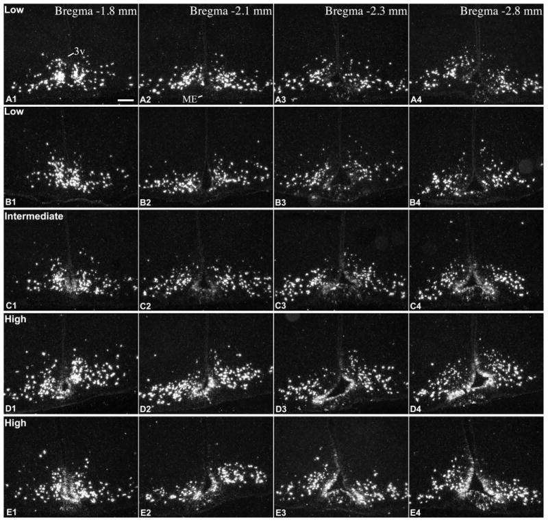
Figure 2.

Radioactive ISH demonstrates Pomc mRNA in the rostral part of the tanycyte region in 5 adult rats with different expression levels in tanycytes of the third ventricle (3v) and non-neuronal cells of the median eminence (ME). (A, B) low, (C) intermediate, (D, E) high Pomc mRNA expression in tanycytes. A, B and D are male, C and E are female rats, between 8-10 weeks of age. Images from the caudal part of the tanycyte region from the same brains are presented in Fig. 3. Scale bar: 200μm.
