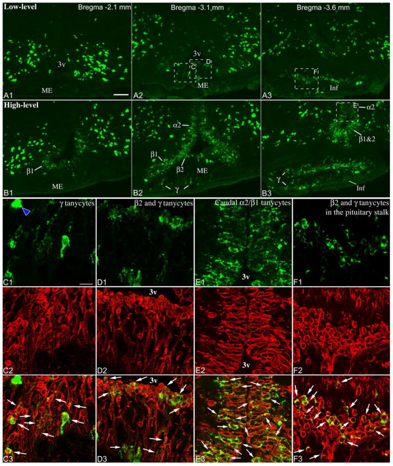
Figure 4.

Low- (A1-3) and high-level (B1-3) Pomc mRNA expression in tanycytes, demonstrated by fluorescent ISH. A1-3 is the same brain as in Fig. 2A, 3A; B1-3 is the same brain as in Fig. 2D, 3D. 3v, third ventricle; α2, α2 tanycytes; β1, β1 tanycytes; β2, β2 tanycytes; γ, γ tanycytes; Inf, infundibular stalk; ME, median eminence. Scale bar: 100μm. (C-F) Confocal images of boxed areas from A2, A3 and B3 show combined Pomc ISH (green) and vimentin immunofluorescence (red). Virtually all non-neuronal Pomc mRNA-expressing cells contain the tanycyte marker vimentin (arrows). Blue arrowhead on C1 points to a Pomc neuron in the median eminence. HuC/D immunofluorescence (not shown) allowed unambiguous identification of Pomc neurons. C, D and E are projections of multiple confocal planes; F shows a single confocal plane. Scale bar: 25μm.
