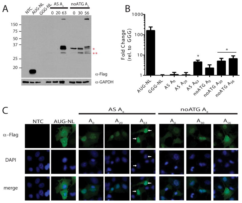
Figure 4. RAN translation from GCC repeats in the alanine reading frame of ASFMR1.
A) western blot against FLAG on lysates from COS-7 cells transfected with indicated ASFMRpolyA reporters. Red asterisks indicate bands generated from initiation 3′ to the repeat site but in the human sequence at a non-canonical start codon. “noATG” indicates the ATG codon in the proline reading frame was absent. B) NanoLuciferase activity from ASFMRpolyA constructs compared to GGG-NL. *=p<0.05 by Fisher’s LSD with Bonferroni correction for individual comparisons to GGG-NL and by ANOVA for repeat length dependent differences among RAN reporters. C) Localization of ASFMRpolyA (green) was primarily nuclear (arrows) compared to AUG-NL which was cytoplasmic. DAPI (blue) was used to counterstain nuclei.

