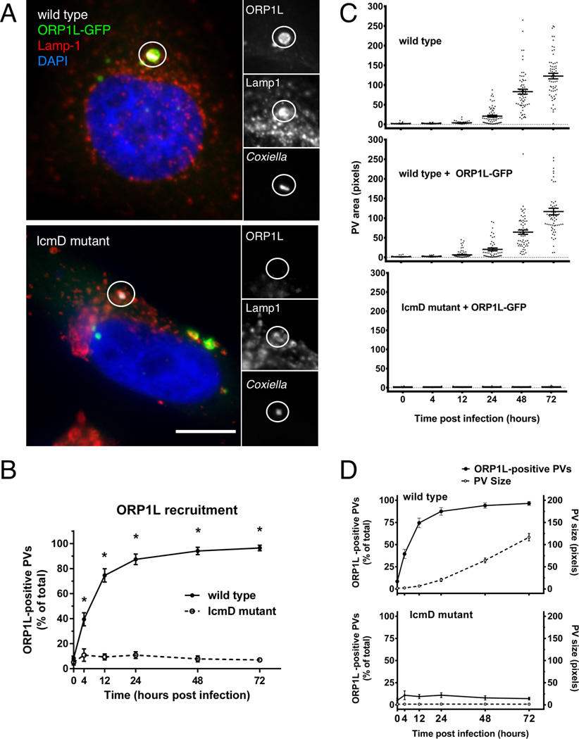
Figure 2. T4BSS-dependent recruitment of ORP1L to the PV membrane.
(A) Light microscopy images of HeLa cells transfected with ORP1L-GFP and then infected with C. burnetii for 2 days. ORP1L-GFP is recruited to the PV of wild type (top) but not IcmD mutant (bottom) bacteria. Green = ORP1L-GFP, Red = Lamp1, Gray = C. burnetii, Blue = DAPI. The PV (circled) is shown magnified with individual fluorescent channels. Scale bar = 10 µm. (B) The number of ORP1L-positive PVs was quantified over a 72 hour time course of infection in HeLa cells infected with wild type (solid) or IcmD mutant (dashed) C. burnetii. Cells were transfected with ORP1L-GFP, infected, stained by immunofluorescence for C. burnetii and Lamp1, and visually scored for the presence or absence of ORP1L on the PV. Each data point represents the average of 3–4 experiments, with 20 PVs counted per experiment. Error bars represent standard error of the mean (SEM). Means compared by unpaired Welch’s t-test. * = p <0.01. (C) Measurements of PV size shows that PVs harboring wild type bacteria expand between 24 and 48 hours, while IcmD mutant PVs do not expand. HeLa cells, untransfected or expressing ORP1L-GFP, were infected with either wild type C. burnetii or the IcmD mutant. At the indicated times, the cells were fixed, stained for C. burnetii and CD63, and the PVs measured using ImageJ. PVs harboring wild type bacteria expanded between 24 and 48 hours, while IcmD PVs did not expand. Shown are individual PV measurements from three separate experiments, with at least 20 PVs per timepoint per experiment. Bars represent mean ± SEM. (D) In the comparison of ORP1L localization compared to PV expansion, ORP1L localizes to the PV 12 hours prior to significant expansion of wild type PVs (top). Error bars represent SEM.

