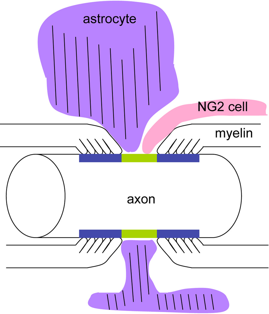
Figure 10.
A scheme depicting a node of Ranvier contacted by an astrocyte (purple) and an NG2 cell (pink). The nodal axolemma is indicated in green and the paranodal membrane in blue. An astrocyte process that contains glial filaments (parallel lines) broadly surrounds the node, whereas an NG2 cell process is more slender and finger-like, coming into contact both at the node and on the myelin paranodal surface.

