Figure 4.
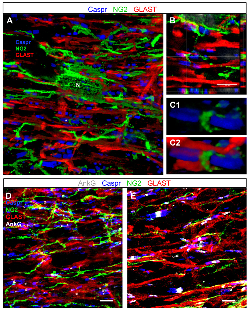
Triple and quadruple immunostaining of P30 optic nerve and thoracic spinal cord
A–C: P30 mouse optic nerve section triple labeled with anti-NG2 (green), GLAST (red), and Caspr (blue) antibodies.
A: A 3D volume rendering showing many Caspr+ paranodal pairs in contact with or flanking glial processes labeled with antibodies to NG2 and GLAST. Several processes from the same NG2 cell (N) are shown to contact different nodes of Ranvier.
B: Single z-plane images in three dimensions show NG2+ and GLAST+ processes positioned directly between Caspr+ paranodes, presumably in close contact with the node (shown with * in A). Image taken from the same z-stack as the volume rendering in panel A.
C1–C2: A 3D volume rendering of the node shown at the crosshairs in B, showing clear insertion of the NG2+ process between the Caspr+ paranodes (C1). The same node is shown in C2 along with the GLAST+ astrocyte processes.
D–E: Quadruple immunostaining of P30 rat optic nerve (D) and P30 mouse thoracic spinal cord white matter (E) sections labeled with AnkG (white), NG2 (green), GLAST (red), and Caspr (blue) antibodies. Images are confocal maximum projections of z-stack images. GLAST+ (astrocyte) and NG2+ cell processes contact the AnkG+ nodes, flanked by Caspr+ paranodes.
Scale bars, 5 µm.

