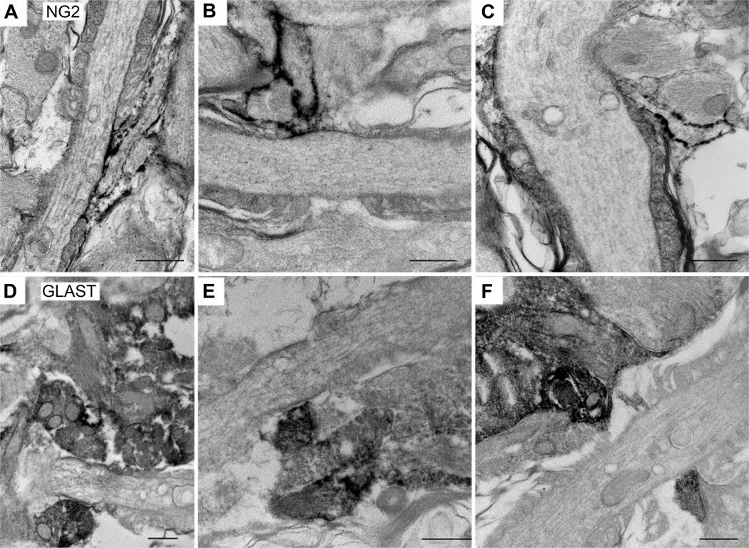
Figure 8.
Pre-embedding immunoelectron microscopy of glial cells at the nodes.
A–C: Immunoperoxidase labeling using rabbit anti-NG2 antibody. A, B: P30 rat optic nerve; C: P30 mouse optic nerve. Slender immunolabeled NG2+ processes insert into the nodes obliquely and are directly apposed to a portion of the nodal axonal membrane.
D–F: Immunoperoxidase labeling using guinea pig anti-GLAST antibody. P30 rat optic nerve. GLAST+ processes appear as broad structures that cover the nodal membrane. In most cases, these processes do not seem to directly contact the axolemma.
Scale bars = 500 nm.

