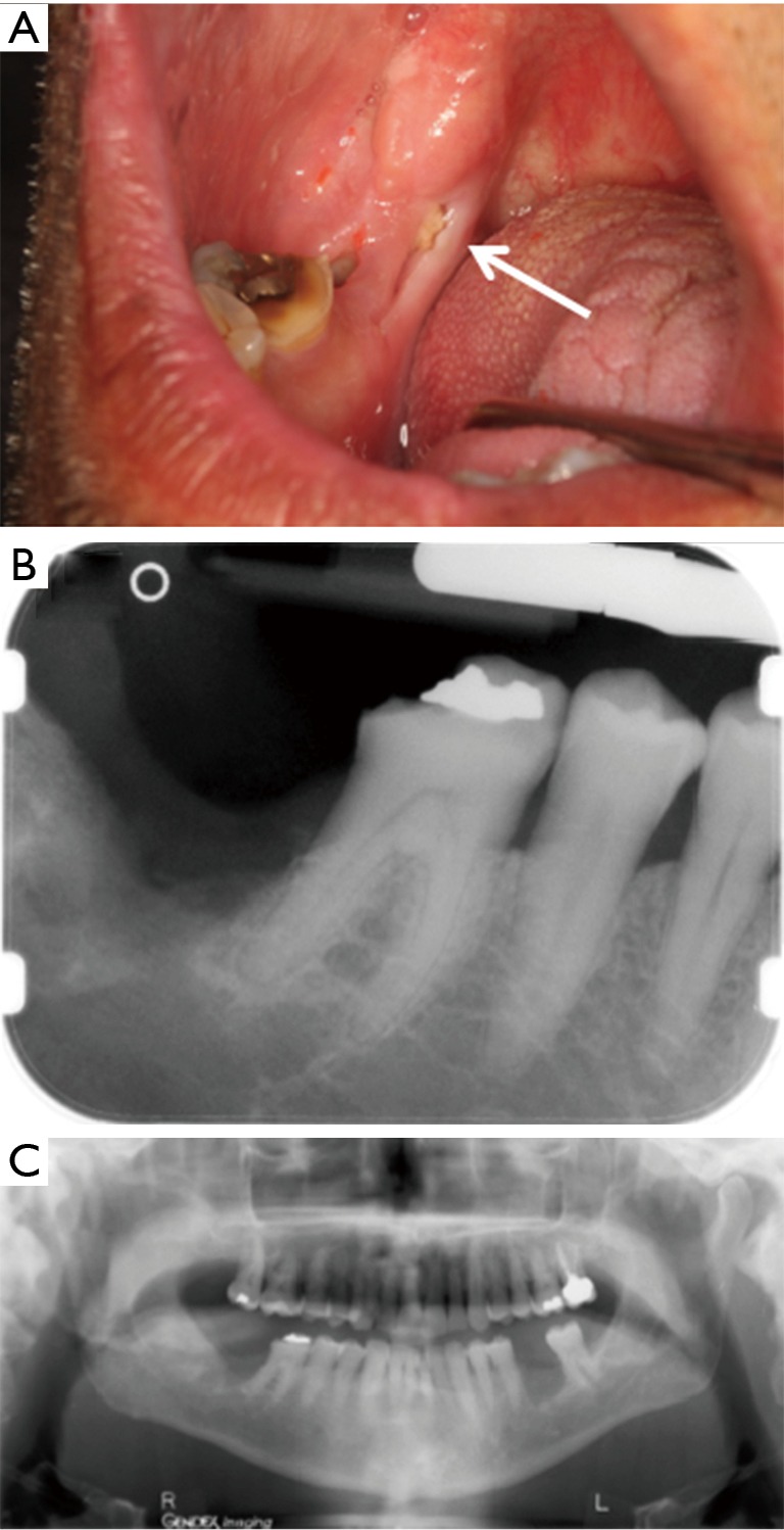
Figure 3.

Case 3. (A) Exposed necrotic bone close to extraction site of right mandibular second and third molars (arrow); (B) periapical radiograph shows ill-defined area of radiolucency distal to mandibular right first molar; (C) panoramic radiograph shows a similar radiolucency but no other lesions.
