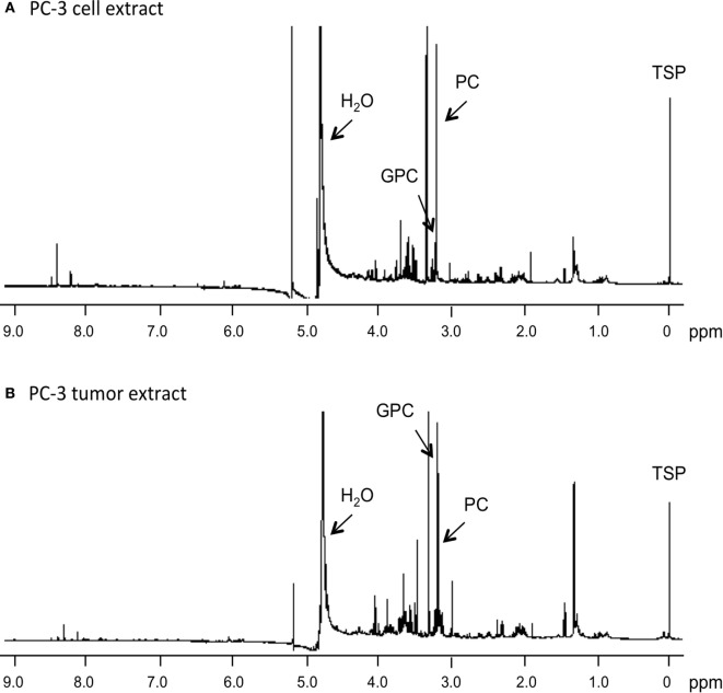
Figure 1.
Representative 1H MR spectra obtained from water-soluble extracts of (A) PC-3 cells (4 × 10⁷ cells) and (B) PC-3 tumor xenograft (0.4 g). Spectra were acquired on a Bruker Avance 11.7 T spectrometer with a 30° flip angle, 10,000 Hz sweep width, 11.2 s repetition time, 32 K block size, and 128 scans. TSP, 3-(trimethylsilyl)propionic 2,2,3,3-d4 acid, an internal standard at 0 ppm; PC, phosphocholine; GPC, glycerophosphocholine; H2O, water signal.

