
FIGURE 4.
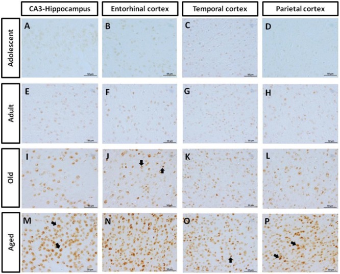
Immunohistochemistry for AT-100 in different brain regions of marmoset. Adolescent animals (A–D) presented a light phosphorylation of tau residues Thr212 and Ser214 in all regions analyzed. Adult subjects (E–H) showed same pattern of immunoreactivity. Old individuals (I–L) had a stronger labeling as AT100-ir appears in cytoplasm, but also as inclusions in the nucleus of the cells. In the aged individuals (M–P) AT100-ir increased in all brain regions of the brain analyzed (cytoplasmic and nuclear inclusions). Inclusions indicated by arrows. Scale bar 50 μm.