. 2016 Nov 30;59(Suppl 1):S1–S4. doi: 10.3345/kjp.2016.59.11.S1

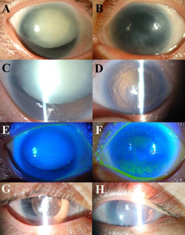
Fig. 3. Ophthalmic examination in 33-year-old subject II-2. Panels A and B are anterior segment photographs of both eyes showing a hypermature cataract in the right eye (A) and corneal opacity in the left eye (B). Panels C and D are slit lamp beam photographs of both eyes showing a dense white cataract and partial existence of an iris in the right eye (C), and a posterior chamber intraocular lens implant, which was inserted following cataract extraction in the left eye (D). Panels E and F show fluorescein dye staining in cobalt blue beam photographs of both eyes with a normal cornea in the right eye (E) and superficial punctate epithelial erosions in the superior and inferior cornea of the left eye (F). Panels G and H are postoperative anterior segment photographs of both eyes.

Fig. 3