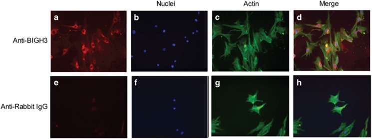
Figure 1.
Immunocytochemistry localization of BIGH3 protein in cultured HRP. Primary HRP were cultured in EBM-2 supplemented with 10% FBS and stained with BIGH3 antibody (red, a) or rabbit IgG (e; negative control) followed by DAPI (nuclei; blue, b and f), phalloidin (actin; green, c and g). In d (a–c merged) shows cytosolic and extracellular locations of BIGH3 protein. Images are shown at × 20 magnification.

