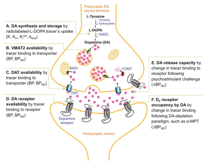
Figure 1. Dopaminergic Imaging Targets.
Schematic of imaging methods used to measure aspects of the dopamine (DA) system in vivo. Graphic depicts progression of DA from synthesis (A), storage (B), to release (E,F), then either reuptake by dopamine transporter (DAT, C) or binding to receptor (D). Imaging targets and related paradigms are described in accompanying text.

