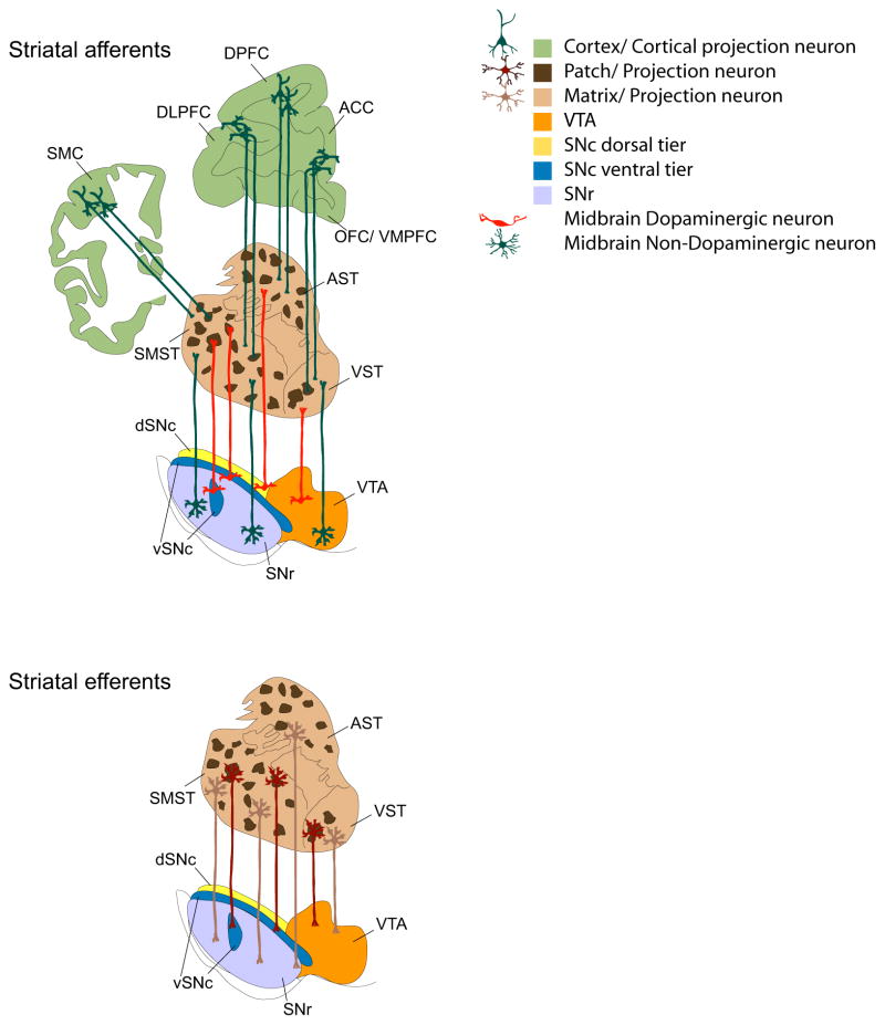
Figure 4. Striatal patch-matrix connectome.
Schematic representation of striatal patch-matrix connectome. Afferents: The cortex topographically projects to the striatum. Within the cortex deeper cortical layers innervate striatal patches (dark brown) whereas the surrounding matrix (light brown) is innervated by superficial cortical layers (light brown). Within the midbrain, the dorsal tier (orange and yellow) innervates the matrix, as do the non-dopaminergic cells (dark green) from the same region. Patch innervation from the midbrain is mostly derived from the ventral tier cell groups (dark blue). Non-dopaminergic (presumably GABAergic) projection neurons within the SNr innervate the striatal matrix complex. Efferents: Striatal patch neurons (maroon) mostly project to ventral tier DA cells. These include both D1 receptor expressing medium spiny neurons and other striatal projection neurons. Striatal projection neurons within the matrix project to both DA and non-dopaminergic populations within the dorsal tier and GABAergic populations in the SNr. See text for further details.

