
Figure 2. Antibodies internalize into murine macrophages and reduce intracellular number of L. monocytogenes.
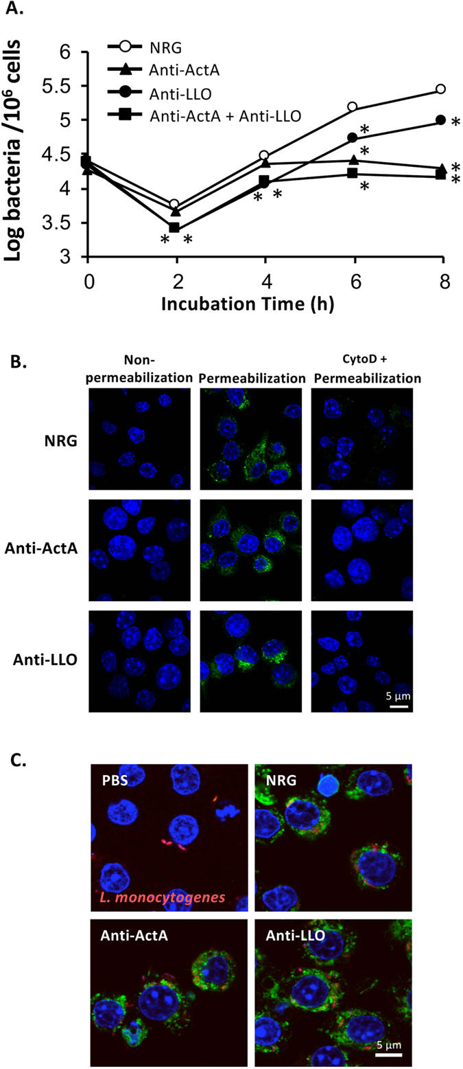
RAW264.7 cells were plated on 24-well culture plates at 1 × 10⁶ cell/well. (A) The cells were treated with 100 μg of the antibody or NRG and infected simultaneously with L. monocytogenes at MOI 10. After incubation for 30 min, the extracellular bacteria were eliminated with 50 μg/ml gentamicin. At each time point, intracellular bacterial number was enumerated (mean ± S.D., n = 9). *P < 0.01. (B) The cells were treated with 100 μg of the antibody or NRG for 2 h. After fixing, immunostaining was performed using AlexaFluor 488-conjugated donkey anti-rabbit IgG. DAPI was used to stain cell nucleus. Left panel: the cells were not permeabilized with Triton X-100, middle and right panels: the cells were permeabilized with Triton X-100, right panel: endocytosis was blocked by cytochalasin D before treatment with the antibody. (C) The cells were treated 100 μg of the antibody or NRG or PBS and infected simultaneously with DsRedEx-labeled L. monocytogenes at MOI 10. After incubation for 30 min, the extracellular bacteria were washed and eliminated with 50 μg/ml gentamicin. After fixation and permeabilization, immunostaining was performed using AlexaFluor 488-conjugated donkey anti-rabbit IgG. DAPI was used to stain cell nucleus.