
Figure 3. Neutralizing activity of the anti-LLO antibody.
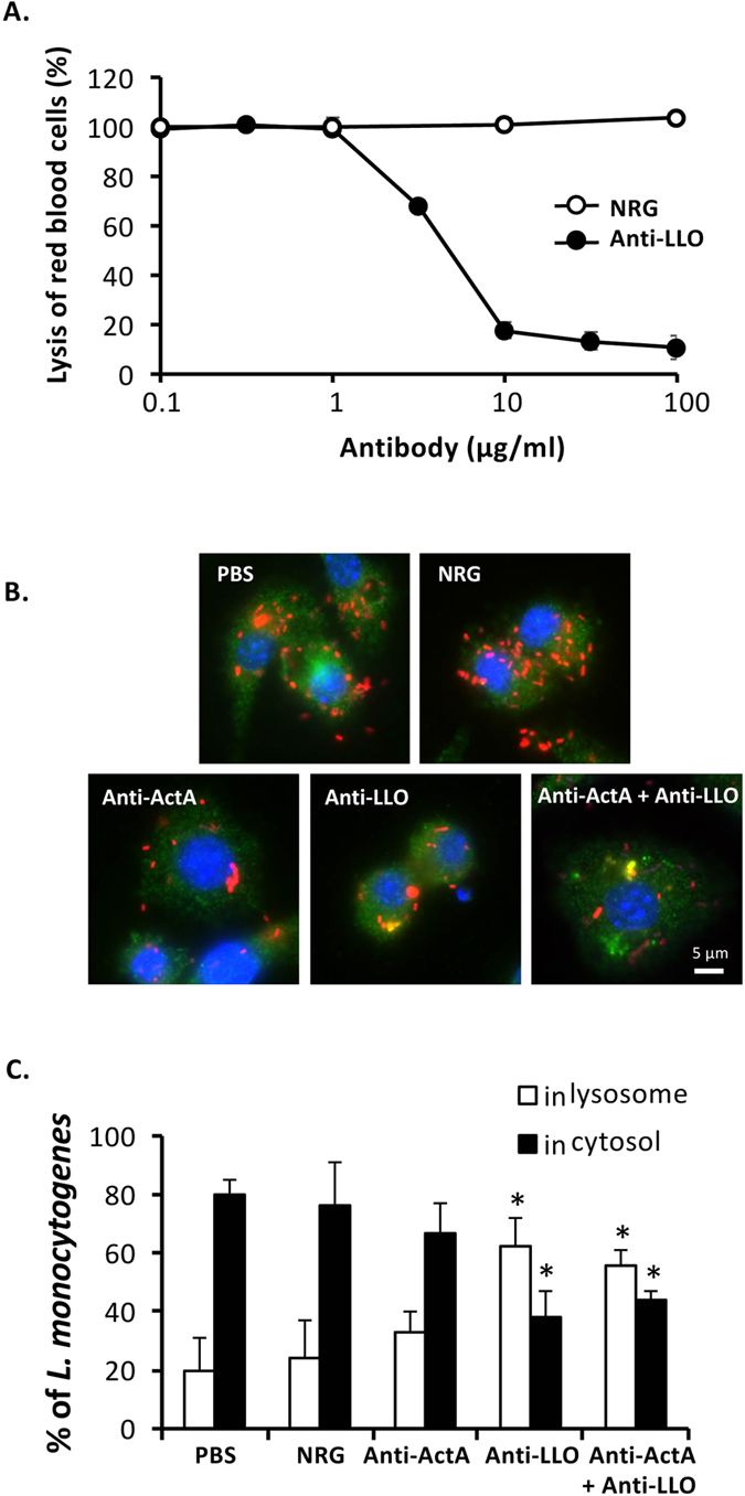
(A) rLLO (1.0 μg/ml) was pre-incubated with anti-LLO antibody or NRG for 1 h at 37 °C prior to incubation with RBC at 37 °C for 45 min. Lysis of RBC was determined at absorbance 541 nm (mean ± S.D., n = 3). Neutralization of hemolytic activity of LLO by the antibody was determined from a reduction of lysis of RBC. (B) RAW264.7 cells were cultivated on sterilized glass slides. The cells were treated with the antibody, NRG or PBS and infected simultaneously with DsRedEx-expressing L. monocytogenes. After incubation for 30 min, the extracellular bacteria were eliminated by gentamicin. At 4 h after infection, the cells were fixed and permeabilized. The cells were stained using mouse monoclonal anti-LAMP1 antibody and AlexaFluor 488-conjugated donkey anti-mouse IgG. DAPI was used to stain cell nucleus. Fluorescent signals were observed under fluorescence microscope. (C) Percentages of L. monocytogenes localized in lysosome and cytoplasm were quantified from at least 100 areas of 3-independent experiments (mean ± S.D.). *P < 0.01.