
Figure 4. Neutralizing activity of the anti-ActA antibody.
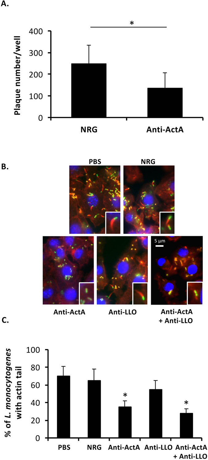
(A) NMuLi cells were treated with the antibody or NRG and infected with simultaneously with L. monocytogenes at MOI 10. Plaques under noble agar were enumerated on day 3 after infection. NRG was used as a control (mean ± S.D., n = 6). *P < 0.05. (B) RAW264.7 cells were cultivated on sterilized glass slides. The cells were treated with the antibody, NRG or PBS and infected simultaneously with YFP-expressing L. monocytogenes. After incubation for 30 min, the extracellular bacteria were eliminated by gentamicin. At 6 h after infection, actin tail was stained using rhodamine-conjugated phalloidin. DAPI was used to stain cell nucleus. (C) Percentage of L. monocytogenes containing actin tail was quantified from at least 100 areas of 3-independent experiments (mean ± S.D.). *P < 0.01.