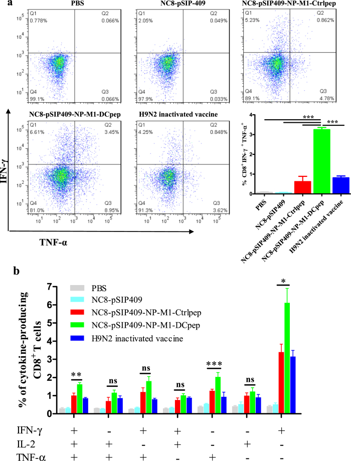
Figure 7. Ag-specific CD8+ T cell cytokine responses after NC8-pSIP409-NP-M1-DCpep vaccination.
(a) Pulmonary CD3+CD8+ T cells from vaccinated mice were assessed by using intracellular cytokine staining for IFN-γ and TNF-α. (b) The frequency of each possible cell population of IFN-γ, TNF-α and IL-2 was evaluated. The data are the mean values ± SEM (n = 5) and were analysed by using a one-way ANOVA, assuming a Gaussian distribution, followed by Dunnett’s post-test and are expressed relative to PBS, NC8-pSIP409 and NC8-pSIP409-NP-M1-Ctrlpep and to the H9N2 inactivated vaccine (**P < 0.01, and ***P < 0.001). The data shown represent one of the three experiments with equivalent results.

