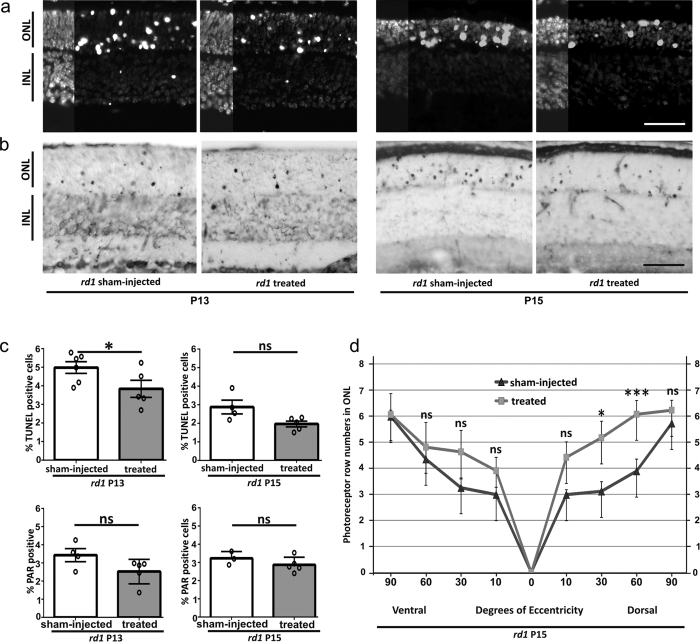
Figure 5. Olaparib protects rd1 photoreceptors in vivo.
After a single intravitreal olaparib injection at P11 rd1 animals where analyzed for cell death (TUNEL assay) and photoreceptor survival at P13 and P15. (a) Retinas from sham-injected rd1 animals display a high number of dying, TUNEL positive cells in the ONL. At P13, in treated animals cell death is reduced, an effect that is still apparent at P15. Note the decrease in the overall size of the rd1 ONL and the number of photoreceptor rows at P15. (b) A subset of rd1 photoreceptors showed a strong immunoreactivity for PARylated proteins, their numbers appeared lower in P13 and P15 treated animals. (c) Top panel: Quantification of TUNEL positive cells in untreated vs. treated animals at P13 and P15. At P13 there were significantly less photoreceptors dying in treated rd1 retina. Bottom panel: Quantification of PAR positive cells in untreated vs. treated animals at P13 and P15. (d) Quantitative analysis of photoreceptor survival along the dorso-ventral axis. While untreated P15 rd1 retina displayed 3-6 photoreceptor rows, olaparib treated rd1 animals had up to two rows more photoreceptors in the mid-periphery, dorsal retina. n(rd1, sham-injected P13) = 6; n(rd1, treated P13) = 5; n(rd1, sham-injected P15) = 4; n(rd1, untreated P15) = 5. Tested by unpaired, two-tailed Student’s t-test. Scale bars in (a and b) are 50 μm.

